72
views
0
recommends
+1 Recommend
0 collections
    0
    shares
      • Record: found
      • Abstract: found
      • Article: found
      Is Open Access

      PINK1 autophosphorylation upon membrane potential dissipation is essential for Parkin recruitment to damaged mitochondria

      research-article

      Read this article at

      Bookmark
          There is no author summary for this article yet. Authors can add summaries to their articles on ScienceOpen to make them more accessible to a non-specialist audience.

          Abstract

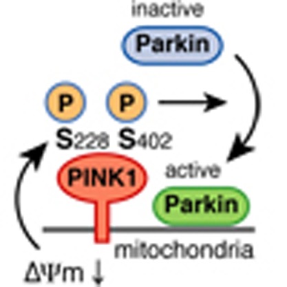
          Dysfunction of PINK1, a mitochondrial Ser/Thr kinase, causes familial Parkinson's disease (PD). Recent studies have revealed that PINK1 is rapidly degraded in healthy mitochondria but accumulates on the membrane potential (ΔΨm)-deficient mitochondria, where it recruits another familial PD gene product, Parkin, to ubiquitylate the damaged mitochondria. Despite extensive study, the mechanism underlying the homeostatic control of PINK1 remains unknown. Here we report that PINK1 is autophosphorylated following a decrease in ΔΨm and that most disease-relevant mutations hinder this event. Mass spectrometric and mutational analyses demonstrate that PINK1 autophosphorylation occurs at Ser228 and Ser402, residues that are structurally clustered together. Importantly, Ala mutation of these sites abolishes autophosphorylation of PINK1 and inhibits Parkin recruitment onto depolarized mitochondria, whereas Asp (phosphorylation-mimic) mutation promotes mitochondrial localization of Parkin even though autophosphorylation was still compromised. We propose that autophosphorylation of Ser228 and Ser402 in PINK1 is essential for efficient mitochondrial localization of Parkin.

          Abstract

          The kinase PINK1 is mutated in Parkinson's disease and accumulates in defective mitochondria, where it recruits Parkin. Here, PINK1 is shown to be autophosphorylated and this is required for the localization of PINK1 to mitochondria with a reduced membrane potential, and for the recruitment of Parkin.

          Related collections

          Most cited references31

          • Record: found
          • Abstract: found
          • Article: not found

          Phosphate-binding tag, a new tool to visualize phosphorylated proteins.

          We introduce two methods for the visualization of phosphorylated proteins using alkoxide-bridged dinuclear metal (i.e. Zn(2+) or Mn(2+)) complexes as novel phosphate-binding tag (Phos-tag) molecules. Both Zn(2+)- and Mn(2+)-Phos-tag molecules preferentially capture phosphomonoester dianions bound to Ser, Thr, and Tyr residues. One method is based on an ECL system using biotin-pendant Zn(2+)-Phos-tag and horseradish peroxidase-conjugated streptavidin. We demonstrate the electroblotting analyses of protein phosphorylation status by the phosphate-selective ECL signals. Another method is based on the mobility shift of phosphorylated proteins in SDS-PAGE with polyacrylamide-bound Mn(2+)-Phos-tag. Phosphorylated proteins in the gel are visualized as slower migration bands compared with corresponding dephosphorylated proteins. We demonstrate the kinase and phosphatase assays by phosphate affinity electrophoresis (Mn(2+)-Phos-tag SDS-PAGE).
            Bookmark
            • Record: found
            • Abstract: found
            • Article: not found

            The conformational plasticity of protein kinases.

            Protein kinases operate in a large number of distinct signaling pathways, where the tight regulation of their catalytic activity is crucial to the development and maintenance of eukaryotic organisms. The catalytic domains of different kinases adopt strikingly similar structures when they are active. By contrast, crystal structures of inactive kinases have revealed a remarkable plasticity in the kinase domain that allows the adoption of distinct conformations in response to interactions with specific regulatory domains or proteins.
              Bookmark
              • Record: found
              • Abstract: found
              • Article: not found

              Regulation of protein kinases; controlling activity through activation segment conformation.

              There are currently at least forty-six unique protein kinase crystal structures, twenty-four of which are available in an active state. Here we examine these structures using a structural bioinformatics approach to understand how the conformation of the activation segment controls kinase activity. Copyright 2004 Cell Press
                Bookmark

                Author and article information

                Journal
                Nat Commun
                Nat Commun
                Nature Communications
                Nature Pub. Group
                2041-1723
                21 August 2012
                : 3
                : 1016
                Affiliations
                [1 ]simpleLaboratory of Protein Metabolism, Tokyo Metropolitan Institute of Medical Science , Setagaya-ku, 156-8506, Japan.
                [2 ]simpleDepartment of Medical Genome Sciences, Graduate School of Frontier Sciences, The University of Tokyo , Kashiwa, Chiba 277-8561, Japan.
                [3 ]simpleDepartment of Life Science, College of Science, Rikkyo University, Toshimaku-ku , Tokyo 171-8501, Japan.
                [4 ]simpleDivision of Disease Proteomics, Institute for Enzyme Research, The University of Tokushima , 770-8503, Japan.
                [5 ]simpleDepartment of Neurology, Juntendo University School of Medicine , Bunkyo-ku, Tokyo 113-8421, Japan.
                [6 ]simpleRIKEN Systems and Structural Biology Center, 1-7-22 Suehiro-cho , Tsurumi-ku, Yokohama 230-0045, Japan.
                [7 ]simpleDepartment of Molecular Biology, Graduate School of Medical Science, Kyushu University , Fukuoka 812-8582, Japan.
                [8 ]simpleProtein Metabolism Project, Tokyo Metropolitan Institute of Medical Science , Setagaya-ku, 156-8506, Japan.
                Author notes
                Article
                ncomms2016
                10.1038/ncomms2016
                3432468
                22910362
                225be4d7-289d-436a-a470-a0aa5c677ecf
                Copyright © 2012, Nature Publishing Group, a division of Macmillan Publishers Limited. All Rights Reserved.

                This work is licensed under a Creative Commons Attribution-NonCommercial-No Derivative Works 3.0 Unported License. To view a copy of this license, visit http://creativecommons.org/licenses/by-nc-nd/3.0/

                History
                : 12 April 2012
                : 20 July 2012
                Categories
                Article

                Uncategorized
                Uncategorized

                Comments

                Comment on this article