4
views
0
recommends
+1 Recommend
0 collections
    0
    shares
      • Record: found
      • Abstract: found
      • Article: found
      Is Open Access

      Recent Advancements in Molecular Therapeutics for Corneal Scar Treatment

      , , , ,
      Cells
      MDPI AG

      Read this article at

      Bookmark
          There is no author summary for this article yet. Authors can add summaries to their articles on ScienceOpen to make them more accessible to a non-specialist audience.

          Abstract

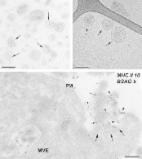
          The process of corneal wound healing is complex and induces scar formation. Corneal scarring is a leading cause of blindness worldwide. The fibrotic healing of a major ocular wound disrupts the highly organized fibrillar collagen arrangement of the corneal stroma, rendering it opaque. The process of regaining this organized extracellular matrix (ECM) arrangement of the stromal layer to restore corneal transparency is complicated. The surface retention capacity of ocular drugs is poor, and there is a large gap between suitable corneal donors and clinical requirements. Therefore, a more efficient way of treating corneal scarring is needed. The eight major classes of interventions targeted as therapeutic tools for healing scarred corneas include those based on exosomes, targeted gene therapy, microRNAs, recombinant viral vectors, histone deacetylase inhibitors, bioactive molecules, growth factors, and nanotechnology. This review highlights the recent advancements in molecular therapeutics to restore a cornea without scarring. It also provides a scope to overcome the limitations of present studies and perform robust clinical research using these strategies.

          Related collections

          Most cited references170

          • Record: found
          • Abstract: found
          • Article: found
          Is Open Access

          Extracellular vesicles: Exosomes, microvesicles, and friends

          Cells release into the extracellular environment diverse types of membrane vesicles of endosomal and plasma membrane origin called exosomes and microvesicles, respectively. These extracellular vesicles (EVs) represent an important mode of intercellular communication by serving as vehicles for transfer between cells of membrane and cytosolic proteins, lipids, and RNA. Deficiencies in our knowledge of the molecular mechanisms for EV formation and lack of methods to interfere with the packaging of cargo or with vesicle release, however, still hamper identification of their physiological relevance in vivo. In this review, we focus on the characterization of EVs and on currently proposed mechanisms for their formation, targeting, and function.
            Bookmark
            • Record: found
            • Abstract: found
            • Article: found
            Is Open Access

            Overview of MicroRNA Biogenesis, Mechanisms of Actions, and Circulation

            MicroRNAs (miRNAs) are a class of non-coding RNAs that play important roles in regulating gene expression. The majority of miRNAs are transcribed from DNA sequences into primary miRNAs and processed into precursor miRNAs, and finally mature miRNAs. In most cases, miRNAs interact with the 3′ untranslated region (3′ UTR) of target mRNAs to induce mRNA degradation and translational repression. However, interaction of miRNAs with other regions, including the 5′ UTR, coding sequence, and gene promoters, have also been reported. Under certain conditions, miRNAs can also activate translation or regulate transcription. The interaction of miRNAs with their target genes is dynamic and dependent on many factors, such as subcellular location of miRNAs, the abundancy of miRNAs and target mRNAs, and the affinity of miRNA-mRNA interactions. miRNAs can be secreted into extracellular fluids and transported to target cells via vesicles, such as exosomes, or by binding to proteins, including Argonautes. Extracellular miRNAs function as chemical messengers to mediate cell-cell communication. In this review, we provide an update on canonical and non-canonical miRNA biogenesis pathways and various mechanisms underlying miRNA-mediated gene regulations. We also summarize the current knowledge of the dynamics of miRNA action and of the secretion, transfer, and uptake of extracellular miRNAs.
              Bookmark
              • Record: found
              • Abstract: found
              • Article: not found

              TGF-beta-induced epithelial to mesenchymal transition.

              During development and in the context of different morphogenetic events, epithelial cells undergo a process called epithelial to mesenchymal transition or transdifferentiation (EMT). In this process, the cells lose their epithelial characteristics, including their polarity and specialized cell-cell contacts, and acquire a migratory behavior, allowing them to move away from their epithelial cell community and to integrate into surrounding tissue, even at remote locations. EMT illustrates the differentiation plasticity during development and is complemented by another process, called mesenchymal to epithelial transition (MET). While being an integral process during development, EMT is also recapitulated under pathological conditions, prominently in fibrosis and in invasion and metastasis of carcinomas. Accordingly, EMT is considered as an important step in tumor progression. TGF-beta signaling has been shown to play an important role in EMT. In fact, adding TGF-beta to epithelial cells in culture is a convenient way to induce EMT in various epithelial cells. Although much less characterized, epithelial plasticity can also be regulated by TGF-beta-related bone morphogenetic proteins (BMPs), and BMPs have been shown to induce EMT or MET depending on the developmental context. In this review, we will discuss the induction of EMT in response to TGF-beta, and focus on the underlying signaling and transcription mechanisms.
                Bookmark

                Author and article information

                Contributors
                Journal
                CELLC6
                Cells
                Cells
                MDPI AG
                2073-4409
                October 2022
                October 21 2022
                : 11
                : 20
                : 3310
                Article
                10.3390/cells11203310
                9600986
                36291182
                14405bae-4a76-496a-988c-b31433cc4906
                © 2022

                https://creativecommons.org/licenses/by/4.0/

                History

                Comments

                Comment on this article