26
views
0
recommends
+1 Recommend
0 collections
    0
    shares
      • Record: found
      • Abstract: found
      • Article: found
      Is Open Access

      microRNA Crosstalk Influences Epithelial-to-Mesenchymal, Endothelial-to-Mesenchymal, and Macrophage-to-Mesenchymal Transitions in the Kidney

      review-article

      Read this article at

      Bookmark
          There is no author summary for this article yet. Authors can add summaries to their articles on ScienceOpen to make them more accessible to a non-specialist audience.

          Abstract

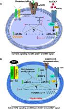
          microRNAs (miRNAs) are small, non-coding nucleotides that regulate diverse biological processes. Altered microRNA biosynthesis or regulation contributes to pathological processes including kidney fibrosis. Kidney fibrosis is characterized by deposition of excess extracellular matrix (ECM), which is caused by infiltration of immune cells, inflammatory cells, altered chemokines, and cytokines as well as activation and accumulation of fibroblasts in the kidney. These activated fibroblasts can arise from epithelial cells via epithelial-to-mesenchymal transition (EMT), from bone marrow-derived M2 phenotype macrophages via macrophage-to-mesenchymal transition (MMT), from endothelial cells via endothelial-to-mesenchymal transition (EndMT), from resident fibroblasts, and from bone marrow-derived monocytes and play a crucial role in fibrotic events. Disrupted microRNA biosynthesis and aberrant regulation contribute to the activation of mesenchymal programs in the kidney. miR-29 regulates the interaction between dipeptidyl peptidase-4 (DPP-4) and integrin β1 and the associated active transforming growth factor β (TGFβ) and pro-EndMT signaling; however, miR-let-7 targets transforming growth factor β receptors (TGFβRs) to inhibit TGFβ signaling. N-acetyl-seryl-aspartyl-lysyl-proline (AcSDKP) is an endogenous anti-fibrotic peptide, which is associated with fibroblast growth factor receptor 1 (FGFR1) phosphorylation and subsequently responsible for the production of miR-let-7. miR-29 and miR-let-7 family clusters participate in crosstalk mechanisms, which are crucial for endothelial cell homeostasis. The physiological level of AcSDKP is vital for the activation of anti-fibrotic mechanisms including restoration of anti-fibrotic microRNA crosstalk and suppression of profibrotic signaling by mitigating DPP-4-associated mesenchymal activation in the epithelial cells, endothelial cells, and M2 phenotype macrophages. The present review highlights recent advancements in the understanding of both the role of microRNAs in the development of kidney disease and their potential as novel therapeutic targets for fibrotic disease states.

          Related collections

          Most cited references141

          • Record: found
          • Abstract: found
          • Article: not found

          Regulation of microRNA function in animals

          Since their serendipitous discovery in nematodes, microRNAs (miRNAs) have emerged as key regulators of biological processes in animals. These small RNAs form complex regulatory networks in cell development, differentiation and homeostasis. Deregulation of miRNA function is associated with an increasing number of human diseases, particularly cancer. Recent discoveries have expanded our understanding of how miRNAs are regulated. Here we review the mechanisms that modulate miRNA activity, their stability and their localization through alternative processing, sequence editing, post-translational modifications of Argonaute proteins, viral factors, transport from the cytoplasm and regulation of miRNA–target interactions. We conclude by discussing intriguing open questions to be answered by future research.
            Bookmark
            • Record: found
            • Abstract: found
            • Article: not found

            Origin and function of myofibroblasts in kidney fibrosis.

            Myofibroblasts are associated with organ fibrosis, but their precise origin and functional role remain unknown. We used multiple genetically engineered mice to track, fate map and ablate cells to determine the source and function of myofibroblasts in kidney fibrosis. Through this comprehensive analysis, we identified that the total pool of myofibroblasts is split, with 50% arising from local resident fibroblasts through proliferation. The nonproliferating myofibroblasts derive through differentiation from bone marrow (35%), the endothelial-to-mesenchymal transition program (10%) and the epithelial-to-mesenchymal transition program (5%). Specific deletion of Tgfbr2 in α-smooth muscle actin (αSMA)(+) cells revealed the importance of this pathway in the recruitment of myofibroblasts through differentiation. Using genetic mouse models and a fate-mapping strategy, we determined that vascular pericytes probably do not contribute to the emergence of myofibroblasts or fibrosis. Our data suggest that targeting diverse pathways is required to substantially inhibit the composite accumulation of myofibroblasts in kidney fibrosis.
              Bookmark
              • Record: found
              • Abstract: found
              • Article: not found

              Fibroblasts in kidney fibrosis emerge via endothelial-to-mesenchymal transition.

              Fibroblasts are key mediators of fibrosis in the kidney and other organs, but their origin during fibrosis is still not completely clear. Activated fibroblasts likely arise from resident quiescent fibroblasts via epithelial-to-mesenchymal transition and from the bone marrow. Here, we demonstrate that endothelial cells also contribute to the emergence of fibroblasts during kidney fibrosis via the process of endothelial-to-mesenchymal transition (EndMT). We examined the contribution of EndMT to renal fibrosis in three mouse models of chronic kidney disease: (1) Unilateral ureteral obstructive nephropathy, (2) streptozotocin-induced diabetic nephropathy, and (3) a model of Alport renal disease. Approximately 30 to 50% of fibroblasts coexpressed the endothelial marker CD31 and markers of fibroblasts and myofibroblasts such as fibroblast specific protein-1 and alpha-smooth muscle actin. Endothelial lineage tracing using Tie2-Cre;R26R-stop-EYFP transgenic mice further confirmed the presence of EndMT-derived fibroblasts. Collectively, our results demonstrate that EndMT contributes to the accumulation of activated fibroblasts and myofibroblasts in kidney fibrosis and suggest that targeting EndMT might have therapeutic potential.
                Bookmark

                Author and article information

                Contributors
                Journal
                Front Pharmacol
                Front Pharmacol
                Front. Pharmacol.
                Frontiers in Pharmacology
                Frontiers Media S.A.
                1663-9812
                16 August 2019
                2019
                : 10
                : 904
                Affiliations
                [1] 1Department of Pediatrics, Yale University School of Medicine , New Haven, CT, United States
                [2] 2Internal Medicine 1, Shimane University Faculty of Medicine , Izumo, Japan
                Author notes

                Edited by: Johan M. Lorenzen, University of Zurich, Switzerland

                Reviewed by: Regalla Kumarswamy, Centre for Cellular and Molecular Biology (CSIR), India; Dan-Qian Chen, Northwest University, China

                *Correspondence: Swayam Prakash Srivastava, swayam.srivastava@ 123456yale.edu ; Keizo Kanasaki, kkanasak@ 123456med.shimane-u.ac.jp ; Julie Goodwin, julie.goodwin@ 123456yale.edu

                This article was submitted to Renal Pharmacology, a section of the journal Frontiers in Pharmacology

                Article
                10.3389/fphar.2019.00904
                6707424
                31474862
                0dbefac0-7c38-4d08-994b-a5c24955c50b
                Copyright © 2019 Srivastava, Hedayat, Kanasaki and Goodwin

                This is an open-access article distributed under the terms of the Creative Commons Attribution License (CC BY). The use, distribution or reproduction in other forums is permitted, provided the original author(s) and the copyright owner(s) are credited and that the original publication in this journal is cited, in accordance with accepted academic practice. No use, distribution or reproduction is permitted which does not comply with these terms.

                History
                : 25 January 2019
                : 18 July 2019
                Page count
                Figures: 6, Tables: 0, Equations: 0, References: 179, Pages: 14, Words: 6690
                Categories
                Pharmacology
                Review

                Pharmacology & Pharmaceutical medicine
                micrornas,diabetic kidney disease,kidney fibrosis,microrna crosstalk,epithelial-to-mesenchymal transition,endothelial-to-mesenchymal transition,macrophage-to-mesenchymal transition

                Comments

                Comment on this article