32
views
0
recommends
+1 Recommend
0 collections
    0
    shares
      • Record: found
      • Abstract: found
      • Article: found
      Is Open Access

      Association of Arch Stiffness with Plantar Impulse Distribution during Walking, Running, and Gait Termination

      research-article

      Read this article at

      Bookmark
          There is no author summary for this article yet. Authors can add summaries to their articles on ScienceOpen to make them more accessible to a non-specialist audience.

          Abstract

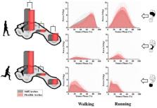
          The purpose of this study was to determine relationships between arch stiffness and relative regional impulse during walking, running, and stopping. A total of 61 asymptomatic male subjects volunteered to participate in the study. All were classified by calculating the arch stiffness index using 3-dimensional foot morphological scanning. Plantar pressure distribution data were collected from participants using a Footscan pressure platform during gait tests that included walking, running, and gait termination. The stiff arches group ( n = 19) and flexible arches group ( n = 17) were included in the following data analysis. The results suggested that subjects with stiffer arches had a larger and smaller percentage of plantar impulse in the forefoot and rearfoot, respectively, than subjects with more flexible arches during walking and running. However, during gait termination, which included planned and unplanned gait stopping, the plantar impulse distribution pattern was found to be reversed. The current findings demonstrate that the distributional changes of plantar loading follow unidirectional transfer between the forefoot and the rearfoot on the plantar longitudinal axis. Moreover, the patterns of impulse distribution are also different based on different gait task mechanisms.

          Related collections

          Most cited references36

          • Record: found
          • Abstract: found
          • Article: not found

          The foot core system: a new paradigm for understanding intrinsic foot muscle function.

          The foot is a complex structure with many articulations and multiple degrees of freedom that play an important role in static posture and dynamic activities. The evolutionary development of the arch of the foot was coincident with the greater demands placed on the foot as humans began to run. The movement and stability of the arch is controlled by intrinsic and extrinsic muscles. However, the intrinsic muscles are largely ignored by clinicians and researchers. As such, these muscles are seldom addressed in rehabilitation programmes. Interventions for foot-related problems are more often directed at externally supporting the foot rather than training these muscles to function as they are designed. In this paper, we propose a novel paradigm for understanding the function of the foot. We begin with an overview of the evolution of the human foot with a focus on the development of the arch. This is followed by a description of the foot intrinsic muscles and their relationship to the extrinsic muscles. We draw the parallels between the small muscles of the trunk region that make up the lumbopelvic core and the intrinsic foot muscles, introducing the concept of the foot core. We then integrate the concept of the foot core into the assessment and treatment of the foot. Finally, we call for an increased awareness of the importance of the foot core stability to normal foot and lower extremity function.
            Bookmark
            • Record: found
            • Abstract: found
            • Article: not found

            Intrinsic foot muscles have the capacity to control deformation of the longitudinal arch.

            The human foot is characterized by a pronounced longitudinal arch (LA) that compresses and recoils in response to external load during locomotion, allowing for storage and return of elastic energy within the passive structures of the arch and contributing to metabolic energy savings. Here, we examine the potential for active muscular contribution to the biomechanics of arch deformation and recoil. We test the hypotheses that activation of the three largest plantar intrinsic foot muscles, abductor hallucis, flexor digitorum and quadratus plantae is associated with muscle stretch in response to external load on the foot and that activation of these muscles (via electrical stimulation) will generate sufficient force to counter the deformation of LA caused by the external load. We found that recruitment of the intrinsic foot muscles increased with increasing load, beyond specific load thresholds. Interestingly, LA deformation and muscle stretch plateaued towards the maximum load of 150% body weight, when muscle activity was greatest. Electrical stimulation of the plantar intrinsic muscles countered the deformation that occurred owing to the application of external load by reducing the length and increasing the height of the LA. These findings demonstrate that these muscles have the capacity to control foot posture and LA stiffness and may provide a buttressing effect during foot loading. This active arch stiffening mechanism may have important implications for how forces are transmitted during locomotion and postural activities as well as consequences for metabolic energy saving.
              Bookmark
              • Record: found
              • Abstract: found
              • Article: not found

              Measurements used to characterize the foot and the medial longitudinal arch: reliability and validity.

              Abnormality in the structure of the medial longitudinal arch of the foot is commonly thought to be a predisposing factor to injury. The purpose of this investigation was to compare the reliability and validity of several measurements used to characterize various aspects of the foot, including the medial longitudinal arch. One hundred two feet (both feet of 51 subjects) were measured to establish a reference database. From this group, a subset of 20 feet (both feet of 10 subjects) was used to determine intertester and intratester reliability. Radiographs of a further subset of 10 feet (right feet of 10 subjects) were used to determine validity. Five foot measurements were taken in 2 stance conditions: 10% of weight bearing and 90% of weight bearing. Intraclass correlation coefficients (ICCs) for intertester and intratester measurements were between.480 and.995. The most reliable method of characterizing arch type in 10% of weight bearing between testers was dividing navicular height by foot length in 10% of weight bearing. However, this measure yielded highly unreliable measurements in 90% of weight bearing. The most valid measurements were navicular height divided by truncated foot length, navicular height divided by foot length in 10% of weight bearing, and navicular height divided by foot length in 90% of weight bearing. Dorsum height at 50% of foot length divided by truncated foot length showed relatively high intertester reliability (ICC=.811 in 10% of weight bearing, ICC=.848 in 90% of weight bearing) and validity (ICC=.844 in 10% of weight bearing, ICC=.851 in 90% of weight bearing). These data suggest that, of the measures tested, the most reliable and valid method of clinically assessing arch height across 10% and 90% of weight bearing was dividing the dorsum height at 50% of foot length by truncated foot length.
                Bookmark

                Author and article information

                Journal
                Int J Environ Res Public Health
                Int J Environ Res Public Health
                ijerph
                International Journal of Environmental Research and Public Health
                MDPI
                1661-7827
                1660-4601
                21 March 2020
                March 2020
                : 17
                : 6
                : 2090
                Affiliations
                [1 ]Faculty of Sports Science, Ningbo University, Ningbo 315211, China; cenxuanzhen@ 123456outlook.com (X.C.); andywutong@ 123456foxmail.com (D.X.)
                [2 ]Department of Sport and Physical Education, Hong Kong Baptist University, Hong Kong, China; jsbaker@ 123456hkbu.edu.hk
                Author notes
                [* ]Correspondence: guyaodong@ 123456nbu.edu.cn ; Tel.: +86-574-8760-0456
                Author information
                https://orcid.org/0000-0002-9093-7897
                https://orcid.org/0000-0003-2187-9440
                Article
                ijerph-17-02090
                10.3390/ijerph17062090
                7143069
                32245235
                8d86ddb1-265f-418b-bf05-5ffd4a3980ab
                © 2020 by the authors.

                Licensee MDPI, Basel, Switzerland. This article is an open access article distributed under the terms and conditions of the Creative Commons Attribution (CC BY) license ( http://creativecommons.org/licenses/by/4.0/).

                History
                : 23 January 2020
                : 20 March 2020
                Categories
                Article

                Public health
                arch stiffness index,unplanned gait termination,gait stop,impulse,arch structure
                Public health
                arch stiffness index, unplanned gait termination, gait stop, impulse, arch structure

                Comments

                Comment on this article