45
views
0
recommends
+1 Recommend
0 collections
    0
    shares
      • Record: found
      • Abstract: found
      • Article: found
      Is Open Access

      Local palmitoylation cycles define activity-regulated postsynaptic subdomains

      research-article

      Read this article at

      Bookmark
          There is no author summary for this article yet. Authors can add summaries to their articles on ScienceOpen to make them more accessible to a non-specialist audience.

          Abstract

          Local palmitoylation machinery has an instructive role in creating activity-responsive PSD-95 nanodomains, which contribute to postsynaptic density (re)organization.

          Abstract

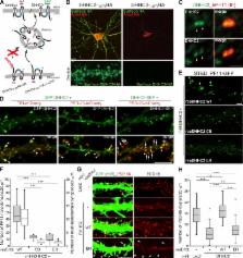
          Distinct PSD-95 clusters are primary landmarks of postsynaptic densities (PSDs), which are specialized membrane regions for synapses. However, the mechanism that defines the locations of PSD-95 clusters and whether or how they are reorganized inside individual dendritic spines remains controversial. Because palmitoylation regulates PSD-95 membrane targeting, we combined a conformation-specific recombinant antibody against palmitoylated PSD-95 with live-cell super-resolution imaging and discovered subsynaptic nanodomains composed of palmitoylated PSD-95 that serve as elementary units of the PSD. PSD-95 in nanodomains underwent continuous de/repalmitoylation cycles driven by local palmitoylating activity, ensuring the maintenance of compartmentalized PSD-95 clusters within individual spines. Plasma membrane targeting of DHHC2 palmitoyltransferase rapidly recruited PSD-95 to the plasma membrane and proved essential for postsynaptic nanodomain formation. Furthermore, changes in synaptic activity rapidly reorganized PSD-95 nano-architecture through plasma membrane–inserted DHHC2. Thus, the first genetically encoded antibody sensitive to palmitoylation reveals an instructive role of local palmitoylation machinery in creating activity-responsive PSD-95 nanodomains, contributing to the PSD (re)organization.

          Related collections

          Most cited references43

          • Record: found
          • Abstract: found
          • Article: not found

          Neurexins induce differentiation of GABA and glutamate postsynaptic specializations via neuroligins.

          Formation of synaptic connections requires alignment of neurotransmitter receptors on postsynaptic dendrites opposite matching transmitter release sites on presynaptic axons. beta-neurexins and neuroligins form a trans-synaptic link at glutamate synapses. We show here that neurexin alone is sufficient to induce glutamate postsynaptic differentiation in contacting dendrites. Surprisingly, neurexin also induces GABA postsynaptic differentiation. Conversely, neuroligins induce presynaptic differentiation in both glutamate and GABA axons. Whereas neuroligins-1, -3, and -4 localize to glutamate postsynaptic sites, neuroligin-2 localizes primarily to GABA synapses. Direct aggregation of neuroligins reveals a linkage of neuroligin-2 to GABA and glutamate postsynaptic proteins, but the other neuroligins only to glutamate postsynaptic proteins. Furthermore, mislocalized expression of neuroligin-2 disperses postsynaptic proteins and disrupts synaptic transmission. Our findings indicate that the neurexin-neuroligin link is a core component mediating both GABAergic and glutamatergic synaptogenesis, and differences in isoform localization and binding affinities may contribute to appropriate differentiation and specificity.
            Bookmark
            • Record: found
            • Abstract: found
            • Article: not found

            Superresolution imaging of chemical synapses in the brain.

            Determination of the molecular architecture of synapses requires nanoscopic image resolution and specific molecular recognition, a task that has so far defied many conventional imaging approaches. Here, we present a superresolution fluorescence imaging method to visualize the molecular architecture of synapses in the brain. Using multicolor, three-dimensional stochastic optical reconstruction microscopy, the distributions of synaptic proteins can be measured with nanometer precision. Furthermore, the wide-field, volumetric imaging method enables high-throughput, quantitative analysis of a large number of synapses from different brain regions. To demonstrate the capabilities of this approach, we have determined the organization of ten protein components of the presynaptic active zone and the postsynaptic density. Variations in synapse morphology, neurotransmitter receptor composition, and receptor distribution were observed both among synapses and across different brain regions. Combination with optogenetics further allowed molecular events associated with synaptic plasticity to be resolved at the single-synapse level. Copyright © 2010 Elsevier Inc. All rights reserved.
              Bookmark
              • Record: found
              • Abstract: found
              • Article: not found

              Neuroligins determine synapse maturation and function.

              Synaptogenesis, the generation and maturation of functional synapses between nerve cells, is an essential step in the development of neuronal networks in the brain. It is thought to be triggered by members of the neuroligin family of postsynaptic cell adhesion proteins, which may form transsynaptic contacts with presynaptic alpha- and beta-neurexins and have been implicated in the etiology of autism. We show that deletion mutant mice lacking neuroligin expression die shortly after birth due to respiratory failure. This respiratory failure is a consequence of reduced GABAergic/glycinergic and glutamatergic synaptic transmission and network activity in brainstem centers that control respiration. However, the density of synaptic contacts is not altered in neuroligin-deficient brains and cultured neurons. Our data show that neuroligins are required for proper synapse maturation and brain function, but not for the initial formation of synaptic contacts.
                Bookmark

                Author and article information

                Journal
                J Cell Biol
                J. Cell Biol
                jcb
                The Journal of Cell Biology
                The Rockefeller University Press
                0021-9525
                1540-8140
                8 July 2013
                : 202
                : 1
                : 145-161
                Affiliations
                [1 ]Division of Membrane Physiology, Department of Cell Physiology, National Institute for Physiological Sciences, Okazaki, Aichi 444-8787, Japan
                [2 ]Centre National de la Recherche Scientifique, Unité Mixte de Recherche 144 and [3 ]Institut Curie Centre de Recherche, 75248 Paris Cedex 05, France
                Author notes
                Correspondence to Masaki Fukata: mfukata@ 123456nips.ac.jp ; or Franck Perez: Franck.Perez@ 123456curie.fr

                F. Perez and M. Fukata contributed equally to this paper.

                O. Vielemeyer’s present address is Drexel University College of Medicine, Philadelphia, PA.

                Article
                201302071
                10.1083/jcb.201302071
                3704990
                23836932
                726ac01e-869c-4078-a0c9-0a9fc377603d
                © 2013 Fukata et al.

                This article is distributed under the terms of an Attribution–Noncommercial–Share Alike–No Mirror Sites license for the first six months after the publication date (see http://www.rupress.org/terms). After six months it is available under a Creative Commons License (Attribution–Noncommercial–Share Alike 3.0 Unported license, as described at http://creativecommons.org/licenses/by-nc-sa/3.0/).

                History
                : 14 February 2013
                : 3 June 2013
                Categories
                Research Articles
                Article

                Cell biology
                Cell biology

                Comments

                Comment on this article