13
views
0
recommends
+1 Recommend
1 collections
    0
    shares
      • Record: found
      • Abstract: found
      • Article: not found

      Tissue distributions of antiviral drugs affect their capabilities of reducing viral loads in COVID-19 treatment

      research-article

      Read this article at

      ScienceOpenPublisherPMC
      Bookmark
          There is no author summary for this article yet. Authors can add summaries to their articles on ScienceOpen to make them more accessible to a non-specialist audience.

          Abstract

          Repurposing of approved antiviral drugs against severe acute respiratory syndrome coronavirus 2 (SARS-CoV-2) is a promising strategy to treat Coronavirus disease 2019 (COVID-19) patients. Previously we reported our hypothesis that the antiviral drugs with high lung distributions might benefit COVID-19 patients by reducing viral loads. So far, chloroquine, lopinavir, hydroxychloroquine, azithromycin, favipiravir, ribavirin, darunavir, remdesivir, and umifenovir have been tested in COVID-19 clinical trials. Here we validated our hypothesis by comparing the pharmacokinetics profiles of these drugs and their capabilities of reducing viral load in clinical trials. According to bulk RNA and single cell RNA sequencing analysis, we found that high expression of both angiotensin converting enzyme 2 (ACE2) and transmembrane Serine Protease 2 (TMPRSS2) makes the lung and intestine vulnerable to SARS-CoV-2. Hydroxychloroquine, chloroquine, and favipiravir, which were highly distributed to the lung, were reported to reduce viral loads in respiratory tract of COVID-19 patients. Conversely, drugs with poor lung distributions, including lopinavir/ritonavir, umifenovir and remdesivir, were insufficient to inhibit viral replication. Lopinavir/ritonavir might inhibit SARS-CoV-2 in the GI tract according to their distribution profiles. We concluded here that the antiviral drugs should be distributed straight to the lung tissue for reducing viral loads in respiratory tract of COVID-19 patients. Additionally, to better evaluate antiviral effects of drugs that target the intestine, the stool samples should also be collected for viral RNA test in the future.

          Graphical abstract

          Related collections

          Most cited references22

          • Record: found
          • Abstract: found
          • Article: not found

          SARS-CoV-2 Cell Entry Depends on ACE2 and TMPRSS2 and Is Blocked by a Clinically Proven Protease Inhibitor

          Summary The recent emergence of the novel, pathogenic SARS-coronavirus 2 (SARS-CoV-2) in China and its rapid national and international spread pose a global health emergency. Cell entry of coronaviruses depends on binding of the viral spike (S) proteins to cellular receptors and on S protein priming by host cell proteases. Unravelling which cellular factors are used by SARS-CoV-2 for entry might provide insights into viral transmission and reveal therapeutic targets. Here, we demonstrate that SARS-CoV-2 uses the SARS-CoV receptor ACE2 for entry and the serine protease TMPRSS2 for S protein priming. A TMPRSS2 inhibitor approved for clinical use blocked entry and might constitute a treatment option. Finally, we show that the sera from convalescent SARS patients cross-neutralized SARS-2-S-driven entry. Our results reveal important commonalities between SARS-CoV-2 and SARS-CoV infection and identify a potential target for antiviral intervention.
            Bookmark
            • Record: found
            • Abstract: found
            • Article: not found

            Virological assessment of hospitalized patients with COVID-2019

            Coronavirus disease 2019 (COVID-19) is an acute infection of the respiratory tract that emerged in late 20191,2. Initial outbreaks in China involved 13.8% of cases with severe courses, and 6.1% of cases with critical courses3. This severe presentation may result from the virus using a virus receptor that is expressed predominantly in the lung2,4; the same receptor tropism is thought to have determined the pathogenicity-but also aided in the control-of severe acute respiratory syndrome (SARS) in 20035. However, there are reports of cases of COVID-19 in which the patient shows mild upper respiratory tract symptoms, which suggests the potential for pre- or oligosymptomatic transmission6-8. There is an urgent need for information on virus replication, immunity and infectivity in specific sites of the body. Here we report a detailed virological analysis of nine cases of COVID-19 that provides proof of active virus replication in tissues of the upper respiratory tract. Pharyngeal virus shedding was very high during the first week of symptoms, with a peak at 7.11 × 108 RNA copies per throat swab on day 4. Infectious virus was readily isolated from samples derived from the throat or lung, but not from stool samples-in spite of high concentrations of virus RNA. Blood and urine samples never yielded virus. Active replication in the throat was confirmed by the presence of viral replicative RNA intermediates in the throat samples. We consistently detected sequence-distinct virus populations in throat and lung samples from one patient, proving independent replication. The shedding of viral RNA from sputum outlasted the end of symptoms. Seroconversion occurred after 7 days in 50% of patients (and by day 14 in all patients), but was not followed by a rapid decline in viral load. COVID-19 can present as a mild illness of the upper respiratory tract. The confirmation of active virus replication in the upper respiratory tract has implications for the containment of COVID-19.
              Bookmark
              • Record: found
              • Abstract: found
              • Article: found
              Is Open Access

              Remdesivir and chloroquine effectively inhibit the recently emerged novel coronavirus (2019-nCoV) in vitro

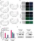
              Dear Editor, In December 2019, a novel pneumonia caused by a previously unknown pathogen emerged in Wuhan, a city of 11 million people in central China. The initial cases were linked to exposures in a seafood market in Wuhan. 1 As of January 27, 2020, the Chinese authorities reported 2835 confirmed cases in mainland China, including 81 deaths. Additionally, 19 confirmed cases were identified in Hong Kong, Macao and Taiwan, and 39 imported cases were identified in Thailand, Japan, South Korea, United States, Vietnam, Singapore, Nepal, France, Australia and Canada. The pathogen was soon identified as a novel coronavirus (2019-nCoV), which is closely related to sever acute respiratory syndrome CoV (SARS-CoV). 2 Currently, there is no specific treatment against the new virus. Therefore, identifying effective antiviral agents to combat the disease is urgently needed. An efficient approach to drug discovery is to test whether the existing antiviral drugs are effective in treating related viral infections. The 2019-nCoV belongs to Betacoronavirus which also contains SARS-CoV and Middle East respiratory syndrome CoV (MERS-CoV). Several drugs, such as ribavirin, interferon, lopinavir-ritonavir, corticosteroids, have been used in patients with SARS or MERS, although the efficacy of some drugs remains controversial. 3 In this study, we evaluated the antiviral efficiency of five FAD-approved drugs including ribavirin, penciclovir, nitazoxanide, nafamostat, chloroquine and two well-known broad-spectrum antiviral drugs remdesivir (GS-5734) and favipiravir (T-705) against a clinical isolate of 2019-nCoV in vitro. Standard assays were carried out to measure the effects of these compounds on the cytotoxicity, virus yield and infection rates of 2019-nCoVs. Firstly, the cytotoxicity of the candidate compounds in Vero E6 cells (ATCC-1586) was determined by the CCK8 assay. Then, Vero E6 cells were infected with nCoV-2019BetaCoV/Wuhan/WIV04/2019 2 at a multiplicity of infection (MOI) of 0.05 in the presence of varying concentrations of the test drugs. DMSO was used in the controls. Efficacies were evaluated by quantification of viral copy numbers in the cell supernatant via quantitative real-time RT-PCR (qRT-PCR) and confirmed with visualization of virus nucleoprotein (NP) expression through immunofluorescence microscopy at 48 h post infection (p.i.) (cytopathic effect was not obvious at this time point of infection). Among the seven tested drugs, high concentrations of three nucleoside analogs including ribavirin (half-maximal effective concentration (EC50) = 109.50 μM, half-cytotoxic concentration (CC50) > 400 μM, selectivity index (SI) > 3.65), penciclovir (EC50 = 95.96 μM, CC50 > 400 μM, SI > 4.17) and favipiravir (EC50 = 61.88 μM, CC50 > 400 μM, SI > 6.46) were required to reduce the viral infection (Fig. 1a and Supplementary information, Fig. S1). However, favipiravir has been shown to be 100% effective in protecting mice against Ebola virus challenge, although its EC50 value in Vero E6 cells was as high as 67 μM, 4 suggesting further in vivo studies are recommended to evaluate this antiviral nucleoside. Nafamostat, a potent inhibitor of MERS-CoV, which prevents membrane fusion, was inhibitive against the 2019-nCoV infection (EC50 = 22.50 μM, CC50 > 100 μM, SI > 4.44). Nitazoxanide, a commercial antiprotozoal agent with an antiviral potential against a broad range of viruses including human and animal coronaviruses, inhibited the 2019-nCoV at a low-micromolar concentration (EC50 = 2.12 μM; CC50 > 35.53 μM; SI > 16.76). Further in vivo evaluation of this drug against 2019-nCoV infection is recommended. Notably, two compounds remdesivir (EC50 = 0.77 μM; CC50 > 100 μM; SI > 129.87) and chloroquine (EC50 = 1.13 μM; CC50 > 100 μM, SI > 88.50) potently blocked virus infection at low-micromolar concentration and showed high SI (Fig. 1a, b). Fig. 1 The antiviral activities of the test drugs against 2019-nCoV in vitro. a Vero E6 cells were infected with 2019-nCoV at an MOI of 0.05 in the treatment of different doses of the indicated antivirals for 48 h. The viral yield in the cell supernatant was then quantified by qRT-PCR. Cytotoxicity of these drugs to Vero E6 cells was measured by CCK-8 assays. The left and right Y-axis of the graphs represent mean % inhibition of virus yield and cytotoxicity of the drugs, respectively. The experiments were done in triplicates. b Immunofluorescence microscopy of virus infection upon treatment of remdesivir and chloroquine. Virus infection and drug treatment were performed as mentioned above. At 48 h p.i., the infected cells were fixed, and then probed with rabbit sera against the NP of a bat SARS-related CoV 2 as the primary antibody and Alexa 488-labeled goat anti-rabbit IgG (1:500; Abcam) as the secondary antibody, respectively. The nuclei were stained with Hoechst dye. Bars, 100 μm. c and d Time-of-addition experiment of remdesivir and chloroquine. For “Full-time” treatment, Vero E6 cells were pre-treated with the drugs for 1 h, and virus was then added to allow attachment for 2 h. Afterwards, the virus–drug mixture was removed, and the cells were cultured with drug-containing medium until the end of the experiment. For “Entry” treatment, the drugs were added to the cells for 1 h before viral attachment, and at 2 h p.i., the virus–drug mixture was replaced with fresh culture medium and maintained till the end of the experiment. For “Post-entry” experiment, drugs were added at 2 h p.i., and maintained until the end of the experiment. For all the experimental groups, cells were infected with 2019-nCoV at an MOI of 0.05, and virus yield in the infected cell supernatants was quantified by qRT-PCR c and NP expression in infected cells was analyzed by Western blot d at 14 h p.i. Remdesivir has been recently recognized as a promising antiviral drug against a wide array of RNA viruses (including SARS/MERS-CoV 5 ) infection in cultured cells, mice and nonhuman primate (NHP) models. It is currently under clinical development for the treatment of Ebola virus infection. 6 Remdesivir is an adenosine analogue, which incorporates into nascent viral RNA chains and results in pre-mature termination. 7 Our time-of-addition assay showed remdesivir functioned at a stage post virus entry (Fig. 1c, d), which is in agreement with its putative anti-viral mechanism as a nucleotide analogue. Warren et al. showed that in NHP model, intravenous administration of 10 mg/kg dose of remdesivir resulted in concomitant persistent levels of its active form in the blood (10 μM) and conferred 100% protection against Ebola virus infection. 7 Our data showed that EC90 value of remdesivir against 2019-nCoV in Vero E6 cells was 1.76 μM, suggesting its working concentration is likely to be achieved in NHP. Our preliminary data (Supplementary information, Fig. S2) showed that remdesivir also inhibited virus infection efficiently in a human cell line (human liver cancer Huh-7 cells), which is sensitive to 2019-nCoV. 2 Chloroquine, a widely-used anti-malarial and autoimmune disease drug, has recently been reported as a potential broad-spectrum antiviral drug. 8,9 Chloroquine is known to block virus infection by increasing endosomal pH required for virus/cell fusion, as well as interfering with the glycosylation of cellular receptors of SARS-CoV. 10 Our time-of-addition assay demonstrated that chloroquine functioned at both entry, and at post-entry stages of the 2019-nCoV infection in Vero E6 cells (Fig. 1c, d). Besides its antiviral activity, chloroquine has an immune-modulating activity, which may synergistically enhance its antiviral effect in vivo. Chloroquine is widely distributed in the whole body, including lung, after oral administration. The EC90 value of chloroquine against the 2019-nCoV in Vero E6 cells was 6.90 μM, which can be clinically achievable as demonstrated in the plasma of rheumatoid arthritis patients who received 500 mg administration. 11 Chloroquine is a cheap and a safe drug that has been used for more than 70 years and, therefore, it is potentially clinically applicable against the 2019-nCoV. Our findings reveal that remdesivir and chloroquine are highly effective in the control of 2019-nCoV infection in vitro. Since these compounds have been used in human patients with a safety track record and shown to be effective against various ailments, we suggest that they should be assessed in human patients suffering from the novel coronavirus disease. Supplementary information Supplementary information, Materials and Figures
                Bookmark

                Author and article information

                Journal
                Eur J Pharmacol
                Eur J Pharmacol
                European Journal of Pharmacology
                Elsevier B.V.
                0014-2999
                1879-0712
                6 October 2020
                15 December 2020
                6 October 2020
                : 889
                : 173634
                Affiliations
                [a ]Center for Translation Medicine Research and Development, Shenzhen Institutes of Advanced Technology, Chinese Academy of Sciences, Shenzhen, 518055, PR China
                [b ]Department of Genetics, Human Genetics Institute of New Jersey, Rutgers University, Piscataway, NJ, 08854, USA
                Author notes
                []Corresponding author. Center for Translation Medicine Research and Development, Shenzhen Institute of Advanced Technology, Chinese Academy of Sciences, Shenzhen, 518055, PR China.
                [∗∗ ]Corresponding author. Department of Genetics, Human Genetics Institute of New Jersey, Rutgers University, Piscataway, NJ, 08854, USA.
                Article
                S0014-2999(20)30726-3 173634
                10.1016/j.ejphar.2020.173634
                7536545
                70d2464c-678a-46d9-a5fa-c9b2b03c8768
                © 2020 Elsevier B.V. All rights reserved.

                Since January 2020 Elsevier has created a COVID-19 resource centre with free information in English and Mandarin on the novel coronavirus COVID-19. The COVID-19 resource centre is hosted on Elsevier Connect, the company's public news and information website. Elsevier hereby grants permission to make all its COVID-19-related research that is available on the COVID-19 resource centre - including this research content - immediately available in PubMed Central and other publicly funded repositories, such as the WHO COVID database with rights for unrestricted research re-use and analyses in any form or by any means with acknowledgement of the original source. These permissions are granted for free by Elsevier for as long as the COVID-19 resource centre remains active.

                History
                : 11 August 2020
                : 29 September 2020
                : 5 October 2020
                Categories
                Full Length Article

                Pharmacology & Pharmaceutical medicine
                coronavirus disease 2019,tissue distribution,antiviral drugs,hydroxychloroquine,chloroquine,favipiravir

                Comments

                Comment on this article