29
views
0
recommends
+1 Recommend
0 collections
    0
    shares
      • Record: found
      • Abstract: found
      • Article: found
      Is Open Access

      Structural modeling of tissue-specific mitochondrial alanyl-tRNA synthetase ( AARS2) defects predicts differential effects on aminoacylation

      research-article

      Read this article at

      Bookmark
          There is no author summary for this article yet. Authors can add summaries to their articles on ScienceOpen to make them more accessible to a non-specialist audience.

          Abstract

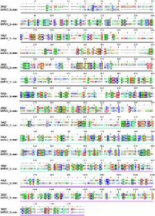
          The accuracy of mitochondrial protein synthesis is dependent on the coordinated action of nuclear-encoded mitochondrial aminoacyl-tRNA synthetases (mtARSs) and the mitochondrial DNA-encoded tRNAs. The recent advances in whole-exome sequencing have revealed the importance of the mtARS proteins for mitochondrial pathophysiology since nearly every nuclear gene for mtARS (out of 19) is now recognized as a disease gene for mitochondrial disease. Typically, defects in each mtARS have been identified in one tissue-specific disease, most commonly affecting the brain, or in one syndrome. However, mutations in the AARS2 gene for mitochondrial alanyl-tRNA synthetase (mtAlaRS) have been reported both in patients with infantile-onset cardiomyopathy and in patients with childhood to adulthood-onset leukoencephalopathy. We present here an investigation of the effects of the described mutations on the structure of the synthetase, in an effort to understand the tissue-specific outcomes of the different mutations. The mtAlaRS differs from the other mtARSs because in addition to the aminoacylation domain, it has a conserved editing domain for deacylating tRNAs that have been mischarged with incorrect amino acids. We show that the cardiomyopathy phenotype results from a single allele, causing an amino acid change R592W in the editing domain of AARS2, whereas the leukodystrophy mutations are located in other domains of the synthetase. Nevertheless, our structural analysis predicts that all mutations reduce the aminoacylation activity of the synthetase, because all mtAlaRS domains contribute to tRNA binding for aminoacylation. According to our model, the cardiomyopathy mutations severely compromise aminoacylation whereas partial activity is retained by the mutation combinations found in the leukodystrophy patients. These predictions provide a hypothesis for the molecular basis of the distinct tissue-specific phenotypic outcomes.

          Related collections

          Most cited references24

          • Record: found
          • Abstract: found
          • Article: not found

          Use of whole-exome sequencing to determine the genetic basis of multiple mitochondrial respiratory chain complex deficiencies.

          Mitochondrial disorders have emerged as a common cause of inherited disease, but their diagnosis remains challenging. Multiple respiratory chain complex defects are particularly difficult to diagnose at the molecular level because of the massive number of nuclear genes potentially involved in intramitochondrial protein synthesis, with many not yet linked to human disease. To determine the molecular basis of multiple respiratory chain complex deficiencies. We studied 53 patients referred to 2 national centers in the United Kingdom and Germany between 2005 and 2012. All had biochemical evidence of multiple respiratory chain complex defects but no primary pathogenic mitochondrial DNA mutation. Whole-exome sequencing was performed using 62-Mb exome enrichment, followed by variant prioritization using bioinformatic prediction tools, variant validation by Sanger sequencing, and segregation of the variant with the disease phenotype in the family. Presumptive causal variants were identified in 28 patients (53%; 95% CI, 39%-67%) and possible causal variants were identified in 4 (8%; 95% CI, 2%-18%). Together these accounted for 32 patients (60% 95% CI, 46%-74%) and involved 18 different genes. These included recurrent mutations in RMND1, AARS2, and MTO1, each on a haplotype background consistent with a shared founder allele, and potential novel mutations in 4 possible mitochondrial disease genes (VARS2, GARS, FLAD1, and PTCD1). Distinguishing clinical features included deafness and renal involvement associated with RMND1 and cardiomyopathy with AARS2 and MTO1. However, atypical clinical features were present in some patients, including normal liver function and Leigh syndrome (subacute necrotizing encephalomyelopathy) seen in association with TRMU mutations and no cardiomyopathy with founder SCO2 mutations. It was not possible to confidently identify the underlying genetic basis in 21 patients (40%; 95% CI, 26%-54%). Exome sequencing enhances the ability to identify potential nuclear gene mutations in patients with biochemically defined defects affecting multiple mitochondrial respiratory chain complexes. Additional study is required in independent patient populations to determine the utility of this approach in comparison with traditional diagnostic methods.
            Bookmark
            • Record: found
            • Abstract: found
            • Article: not found

            Making proteins in the powerhouse.

            Understanding regulation of mitochondrial DNA (mtDNA) expression is of considerable interest given that mitochondrial dysfunction is important in human pathology and aging. Similar to the situation in bacteria, there is no compartmentalization between transcription and translation in mitochondria; hence, both processes are likely to have a direct molecular crosstalk. Accumulating evidence suggests that there are important mechanisms for regulation of mammalian mtDNA expression at the posttranscriptional level. Regulation of mRNA maturation, mRNA stability, translational coordination, ribosomal biogenesis, and translation itself all form the basis for controlling oxidative phosphorylation capacity. Consequently, a wide variety of inherited human mitochondrial diseases are caused by mutations of nuclear genes regulating various aspects of mitochondrial translation. Furthermore, mutations of mtDNA, associated with human disease and aging, often affect tRNA genes critical for mitochondrial translation. Recent advances in molecular understanding of mitochondrial translation regulation will most likely provide novel avenues for modulating mitochondrial function for treating human disease. Copyright © 2014 Elsevier Inc. All rights reserved.
              Bookmark
              • Record: found
              • Abstract: found
              • Article: not found
              Is Open Access

              Mutations of the mitochondrial-tRNA modifier MTO1 cause hypertrophic cardiomyopathy and lactic acidosis.

              Dysfunction of mitochondrial respiration is an increasingly recognized cause of isolated hypertrophic cardiomyopathy. To gain insight into the genetic origin of this condition, we used next-generation exome sequencing to identify mutations in MTO1, which encodes mitochondrial translation optimization 1. Two affected siblings carried a maternal c.1858dup (p.Arg620Lysfs(∗)8) frameshift and a paternal c.1282G>A (p.Ala428Thr) missense mutation. A third unrelated individual was homozygous for the latter change. In both humans and yeast, MTO1 increases the accuracy and efficiency of mtDNA translation by catalyzing the 5-carboxymethylaminomethylation of the wobble uridine base in three mitochondrial tRNAs (mt-tRNAs). Accordingly, mutant muscle and fibroblasts showed variably combined reduction in mtDNA-dependent respiratory chain activities. Reduced respiration in mutant cells was corrected by expressing a wild-type MTO1 cDNA. Conversely, defective respiration of a yeast mto1Δ strain failed to be corrected by an Mto1(Pro622∗) variant, equivalent to human MTO1(Arg620Lysfs∗8), whereas incomplete correction was achieved by an Mto1(Ala431Thr) variant, corresponding to human MTO1(Ala428Thr). The respiratory yeast phenotype was dramatically worsened in stress conditions and in the presence of a paromomycin-resistant (P(R)) mitochondrial rRNA mutation. Lastly, in vivo mtDNA translation was impaired in the mutant yeast strains. Copyright © 2012 The American Society of Human Genetics. Published by Elsevier Inc. All rights reserved.
                Bookmark

                Author and article information

                Contributors
                Journal
                Front Genet
                Front Genet
                Front. Genet.
                Frontiers in Genetics
                Frontiers Media S.A.
                1664-8021
                06 February 2015
                2015
                : 6
                : 21
                Affiliations
                [1] 1Research Programs Unit, Molecular Neurology, Biomedicum Helsinki, University of Helsinki Helsinki, Finland
                [2] 2Department of Clinical Chemistry, University of Gothenburg, Sahlgrenska University Hospital Gothenburg, Sweden
                [3] 3Department of Pediatrics, Queen Silvia Children’s Hospital, University of Gothenburg Gothenburg, Sweden
                [4] 4Institute of Genetic Medicine, Wellcome Trust Centre for Mitochondrial Research, Newcastle University Newcastle upon Tyne, UK
                [5] 5Institute of Neuroscience, Wellcome Trust Centre for Mitochondrial Research, Newcastle University Newcastle upon Tyne, UK
                [6] 6Department of Neuropediatrics, Developmental Neurology and Social Pediatrics, University of Essen Essen, Germany
                [7] 7Murdoch Childrens Research Institute, Royal Childrens Hospital and Department of Paediatrics, University of Melbourne Melbourne, VIC, Australia
                [8] 8Department of Neurology, Helsinki University Central Hospital Helsinki, Finland
                [9] 9Department of Chemistry, Carleton College Northfield, MN, USA
                [10] 10Department of Medical Genetics, Haartman Institute, University of Helsinki Helsinki, Finland
                Author notes

                Edited by: Daniele Ghezzi, Istituto Neurologico Carlo Besta, Italy

                Reviewed by: Lluís Ribas De Pouplana, IRB Barcelona, Spain; Michal Minczuk, Medical Research Council, UK; Daria Diodato, Istituto Neurologico Carlo Besta, Italy

                *Correspondence: Henna Tyynismaa, Research Programs Unit, Molecular Neurology, Biomedicum Helsinki, University of Helsinki, r.C520A, Haartmaninkatu 8, 00290 Helsinki, Finland e-mail: henna.tyynismaa@ 123456helsinki.fi

                This article was submitted to Genetic Disorders, a section of the journal Frontiers in Genetics.

                Article
                10.3389/fgene.2015.00021
                4319469
                25705216
                430e1bd6-1dfb-4899-9b9c-58458a27f9bb
                Copyright © 2015 Euro, Konovalova, Asin-Cayuela, Tulinius, Griffin, Horvath, Taylor, Chinnery, Schara, Thorburn, Suomalainen, Chihade and Tyynismaa.

                This is an open-access article distributed under the terms of the Creative Commons Attribution License (CC BY). The use, distribution or reproduction in other forums is permitted, provided the original author(s) or licensor are credited and that the original publication in this journal is cited, in accordance with accepted academic practice. No use, distribution or reproduction is permitted which does not comply with these terms.

                History
                : 28 November 2014
                : 15 January 2015
                Page count
                Figures: 8, Tables: 3, Equations: 0, References: 36, Pages: 12, Words: 0
                Categories
                Genetics
                Original Research Article

                Genetics
                mitochondrial disease,aminoacyl-trna synthetases,alanyl-trna synthetase,tissue-specificity,structural modeling

                Comments

                Comment on this article