10
views
0
recommends
+1 Recommend
0 collections
    0
    shares
      • Record: found
      • Abstract: found
      • Article: found
      Is Open Access

      Lateral alveolar ridge augmentation procedure using subperiosteal tunneling technique: a pilot study

      research-article

      Read this article at

      Bookmark
          There is no author summary for this article yet. Authors can add summaries to their articles on ScienceOpen to make them more accessible to a non-specialist audience.

          Abstract

          Background

          In this research article, we evaluate the use of sub-periosteal tunneling (tunnel technique) combined with alloplastic in situ hardening biphasic calcium phosphate (BCP, a compound of β-tricalcium phosphate and hydroxyapatite) bone graft for lateral augmentation of a deficient alveolar ridge.

          Methods

          A total of 9 patients with deficient mandibular alveolar ridges were included in the present pilot study. Ten lateral ridge augmentation were carried out using the sub-periosteal tunneling technique, including a bilateral procedure in one patient. The increase in ridge width was assessed using CBCT evaluation of the ridge preoperatively and at 4 months postoperatively. Histological assessment of the quality of bone formation was also carried out with bone cores obtained at the implant placement re-entry in one patient.

          Results

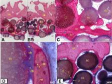
          The mean bucco-lingual ridge width increased in average from 4.17 ± 0.99 mm to 8.56 ± 1.93 mm after lateral bone augmentation with easy-graft CRYSTAL using the tunneling technique. The gain in ridge width was statistically highly significant ( p = 0.0019). Histomorphometric assessment of two bone cores obtained at the time of implant placement from one patient revealed 27.6% new bone and an overall mineralized fraction of 72.3% in the grafted area 4 months after the bone grafting was carried out.

          Conclusions

          Within the limits of this pilot study, it can be concluded that sub-periosteal tunneling technique using in situ hardening biphasic calcium phosphate is a valuable option for lateral ridge augmentation to allow implant placement in deficient alveolar ridges. Further prospective randomized clinical trials will be necessary to assess its performance in comparison to conventional ridge augmentation procedures.

          Related collections

          Most cited references28

          • Record: found
          • Abstract: found
          • Article: not found

          Alveolar bone dimensional changes of post-extraction sockets in humans: a systematic review.

          To review the literature to assess the amount of change in height and width of the residual ridge after tooth extraction. MEDLINE-PubMed and the Cochrane Central register of controlled trials (CENTRAL) were searched through up to March 2009. Appropriate studies which data reported concerning the dimensional changes in alveolar height and width after tooth extraction were included. Approximal height change, mid-buccal change, mid-crestal change, mid-lingual change, Alveolar width change and socket fill were selected as outcome variables. Mean values and if available standard deviations were extracted. Weighted mean changes were calculated. Independent screening of the titles and abstracts of 1244 MEDLINE-PubMed and 106 Cochrane papers resulted in 12 publications that met the eligibility criteria. The reduction in width of the alveolar ridges was 3.87 mm. The mean clinical mid-buccal height loss was 1.67 mm. The mean crestal height change as assessed on the radiographs was 1.53 mm. Socket fill in height as measured relative to the original socket floor was on an average 2.57 mm. During the post-extraction healing period, the weighted mean changes as based on the data derived from the individual selected studies show the clinical loss in width to be greater than the loss in height, assessed both clinically as well as radiographically.
            Bookmark
            • Record: found
            • Abstract: found
            • Article: not found

            Ridge alterations following implant placement in fresh extraction sockets: an experimental study in the dog.

            To study dimensional alterations of the alveolar ridge that occurred following implant placement in fresh extraction sockets. Five beagle dogs were included in the study. In both quadrants of the mandible, incisions were made in the crevice region of the third and fourth pre-molars. Buccal and minute lingual full-thickness flaps were elevated. The mesial root of the four pre-molars root was filled and the teeth were hemi-sected. Following flap elevation in (3)P(3) and (4)P(4) regions, the distal roots were removed. In the right jaw quadrants, implants with a sand blasted and acid etched (SLA) surface were placed in the fresh extraction sockets, while in the left jaws the corresponding sockets were left for spontaneous healing. The mesial roots were retained as surgical control teeth. After 3 months, the animals were examined clinically, sacrificed and tissue blocks containing the implant sites, the adjacent tooth sites (mesial root) and the edentulous socket sites were dissected, prepared for ground sectioning and examined in the microscope. At implant sites, the level of bone-to-implant contact (BC) was located 2.6+/-0.4 mm (buccal aspect) and 0.2+/-0.5 mm (lingual aspect) apical of the SLA level. At the edentulous sites, the mean vertical distance (V) between the marginal termination of the buccal and lingual bone walls was 2.2+/-0.9 mm. At the surgically treated tooth sites, the mean amount of attachment loss was 0.5+/-0.5 mm (buccal) and 0.2+/-0.3 mm (lingual). Marked dimensional alterations had occurred in the edentulous ridge after 3 months of healing following the extraction of the distal root of mandibular pre-molars. The placement of an implant in the fresh extraction site obviously failed to prevent the re-modelling that occurred in the walls of the socket. The resulting height of the buccal and lingual walls at 3 months was similar at implants and edentulous sites and vertical bone loss was more pronounced at the buccal than at the lingual aspect of the ridge. It is suggested that the resorption of the socket walls that occurs following tooth removal must be considered in conjunction with implant placement in fresh extraction sockets.
              Bookmark
              • Record: found
              • Abstract: found
              • Article: not found

              Bone healing and graft resorption of autograft, anorganic bovine bone and beta-tricalcium phosphate. A histologic and histomorphometric study in the mandibles of minipigs.

              The purpose was to qualitatively and quantitatively compare the bone formation and graft resorption of two different bone substitutes used in both orthopedic and oral surgery, with autogenous bone as a positive control. Three standardized bone defects were prepared in both mandibular angles of 12 adult minipigs. The defects were grafted with either autograft, anorganic bovine bone (ABB), or synthetic beta-tricalcium phosphate (beta-TCP). Sacrifice was performed after 1, 2, 4, and 8 weeks for histologic and histomorphometric analysis. At 2 weeks, more new bone formation was seen in defects filled with autograft than with ABB (P approximately 0.0005) and beta-TCP (P approximately 0.002). After 4 weeks, there was no significant difference between beta-TCP and the two other materials. Defects grafted with ABB still exhibited less bone formation as compared with autograft (P approximately 0.004). At 8 weeks, more bone formation was observed in defects grafted with autograft (P approximately 0.003) and beta-TCP (P approximately 0.00004) than with ABB. No difference could be demonstrated between beta-TCP and autograft. beta-TCP resorbed almost completely over 8 weeks, whereas ABB remained stable. Both bone substitutes seemed to decelerate bone regeneration in the early healing phase as compared with autograft. All defects ultimately regenerated with newly formed bone and a developing bone marrow. The grafting materials showed complete osseous integration. Both bone substitutes may have a place in reconstructive surgery where different clinical indications require differences in biodegradability.
                Bookmark

                Author and article information

                Contributors
                kakar_ashish@yahoo.com
                kanupriya100@yahoo.co.in
                principalydc@yenepoya.edu.in
                annette.lindner@uniklinic-freiburg.de
                h-nagursky@t-online.de
                drgauravjain1@gmail.com
                draditya@mahajanimaging.com
                Journal
                Maxillofac Plast Reconstr Surg
                Maxillofac Plast Reconstr Surg
                Maxillofacial Plastic and Reconstructive Surgery
                Springer Berlin Heidelberg (Berlin/Heidelberg )
                2288-8101
                2288-8586
                25 February 2018
                25 February 2018
                December 2018
                : 40
                : 1
                : 3
                Affiliations
                [1 ]ISNI 0000 0004 1767 7704, GRID grid.413027.3, Yenepoya University Dental College, ; University Road, Mangalore, 575018 India
                [2 ]Private Practice, New Delhi, India
                [3 ]GRID grid.5963.9, Institute for Clinical Chemistry and Laboratory Medicine, Medical Center, , University of Freiburg, ; HugstetterStrasse 55, 79106 Freiburg, Germany
                [4 ]ISNI 0000 0004 1804 700X, GRID grid.414612.4, Dental Surgery, , Indraprastha Apollo Hospitals, ; New Delhi, India
                [5 ]Mahajan Imaging Center, New Delhi, India
                Article
                142
                10.1186/s40902-018-0142-8
                6328851
                39c3bb2c-397b-4f75-8a54-b908207565e9
                © The Author(s). 2018

                Open AccessThis article is distributed under the terms of the Creative Commons Attribution 4.0 International License ( http://creativecommons.org/licenses/by/4.0/), which permits unrestricted use, distribution, and reproduction in any medium, provided you give appropriate credit to the original author(s) and the source, provide a link to the Creative Commons license, and indicate if changes were made.

                History
                : 16 August 2017
                : 18 January 2018
                Funding
                Funded by: Sunstar Suisse SA
                Categories
                Research
                Custom metadata
                © The Author(s) 2018

                alveolar,ridge,augmentation,subperiosteal,tunneling,alloplastic
                alveolar, ridge, augmentation, subperiosteal, tunneling, alloplastic

                Comments

                Comment on this article