35
views
0
recommends
+1 Recommend
0 collections
    0
    shares
      • Record: found
      • Abstract: found
      • Article: found
      Is Open Access

      Wnt ligands from the embryonic surface ectoderm regulate ‘bimetallic strip’ optic cup morphogenesis in mouse

      research-article

      Read this article at

      Bookmark
          There is no author summary for this article yet. Authors can add summaries to their articles on ScienceOpen to make them more accessible to a non-specialist audience.

          Abstract

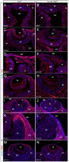
          The Wnt/β-catenin response pathway is central to many developmental processes. Here, we assessed the role of Wnt signaling in early eye development using the mouse as a model system. We showed that the surface ectoderm region that includes the lens placode expressed 12 out of 19 possible Wnt ligands. When these activities were suppressed by conditional deletion of wntless ( Le-cre; Wls fl/fl ) there were dramatic consequences that included a saucer-shaped optic cup, ventral coloboma, and a deficiency of periocular mesenchyme. This phenotype shared features with that produced when the Wnt/β-catenin pathway co-receptor Lrp6 is mutated or when retinoic acid (RA) signaling in the eye is compromised. Consistent with this, microarray and cell fate marker analysis identified a series of expression changes in genes known to be regulated by RA or by the Wnt/β-catenin pathway. Using pathway reporters, we showed that Wnt ligands from the surface ectoderm directly or indirectly elicit a Wnt/β-catenin response in retinal pigment epithelium (RPE) progenitors near the optic cup rim. In Le-cre; Wls fl/fl mice, the numbers of RPE cells are reduced and this can explain, using the principle of the bimetallic strip, the curvature of the optic cup. These data thus establish a novel hypothesis to explain how differential cell numbers in a bilayered epithelium can lead to shape change.

          Abstract

          Summary: During optic cup morphogenesis, Wnt ligands expressed in the surface ectoderm control cell proliferation in the retinal pigmented epithelium, and thus influence bending of the neural retina.

          Related collections

          Most cited references58

          • Record: found
          • Abstract: found
          • Article: not found

          Multiple roles for activated LEF/TCF transcription complexes during hair follicle development and differentiation.

          LEF/TCF DNA-binding proteins act in concert with activated beta -catenin, the product of Wnt signaling, to transactivate downstream target genes. To probe the role of activated LEF/TCF transcription factor complexes in hair follicle morphogenesis and differentiation, we engineered mice harboring TOPGAL, a beta -galactosidase gene under the control of a LEF/TCF and beta -catenin inducible promoter. In mice, TOPGAL expression was directly stimulated by a stabilized form of beta -catenin, but was also dependent upon LEF1/TCF3 in skin. During embryogenesis, TOPGAL activation occurred transiently in a subset of LEF1-positive cells of pluripotent ectoderm and underlying mesenchyme. Downgrowth of initiated follicles proceeded in the absence of detectable TOPGAL expression, even though LEF1 was still expressed. While proliferative matrix cells expressed the highest levels of Lef1 mRNAs, LEF1 concentrated in the precursor cells to the hair shaft, where TOPGAL expression was co-induced with hair-specific keratin genes containing LEF/TCF-binding motifs. LEF1 and TOPGAL expression ceased during catagen and telogen, but reappeared at the start of the postnatal hair cycle, concomitant with precortex formation. In contrast to hair shaft precursor cells, postnatal outer root sheath expressed TCF3, but not TOPGAL. TCF3 was also expressed in the putative follicle stem cells, and while TOPGAL was generally silent in this compartment, it was stimulated at the start of the hair cycle in a fashion that appeared to be dependent upon stabilization of beta -catenin. Taken together, our findings demonstrate that LEF1/TCF3 is necessary but not sufficient for TOPGAL activation, revealing the existence of positive and negative regulators of these factors in the skin. Furthermore, our findings unveil the importance of activated LEF/TCF complexes at distinct times in hair development and cycling when changes in cell fate and differentiation commitments take place.
            Bookmark
            • Record: found
            • Abstract: found
            • Article: not found

            Z/EG, a double reporter mouse line that expresses enhanced green fluorescent protein upon Cre-mediated excision.

            The Cre/loxP system has become an important tool in designing postintegrational switch mechanisms for transgenes in mice. The power and spectrum of application of this system depends on transgenic mouse lines that provide Cre recombinase activity with a defined cell type-, tissue-, or developmental stage-specificity. We have developed a novel mouse line that acts as a Cre reporter. The mice, designated Z/EG (lacZ/EGFP), express lacZ throughout embryonic development and adult stages. Cre excision, however, removes the lacZ gene, which activates expression of the second reporter, enhanced green fluorescent protein. We have found that the double-reporter Z/EG line is able to indicate the occurrence of Cre excision from early embryonic to adult lineages. The advantage of the Z/EG line is that Cre-mediated excision can be monitored in live samples and that live cells with Cre-mediated excision can be isolated using a single-step FACS. It will be a valuable reagent for the increasing number of investigators taking advantage of the powerful tools provided by the Cre/loxP site-specific recombinase system.
              Bookmark
              • Record: found
              • Abstract: found
              • Article: not found

              Specialized filopodia direct long-range transport of Shh during vertebrate tissue patterning

              The ability of signaling proteins to traverse tissues containing tightly packed cells is of fundamental importance for cell specification and tissue development, however, how this is achieved at a cellular level remains poorly understood 1 . For over a century, the vertebrate limb bud has served as a paradigm to study cell signaling during embryonic development 2 . Here we optimize single cell real-time imaging to delineate the cellular mechanisms for how signaling proteins, such as Sonic Hedgehog (Shh), that possess membrane-bound covalent lipid modifications transverse long distances within the limb bud in vivo. By directly imaging Shh ligand production under native regulatory control, our findings show that Shh is unexpectedly produced in the form of a particle that remains associated with the cell via long cytoplasmic extensions that span several cell diameters. We show that these cellular extensions are a specialized class of actin-based filopodia with novel cytoskeletal features that have not been previously described. Strikingly, particles containing Shh traffic along these extensions with a net anterograde movement within the field of Shh cell signaling. We further show that in Shh responding cells specific subsets of Shh co-receptors, including Cdo and Boc, actively distribute and co-localize in specific micro-domains within filopodial extensions, far from the cell body. Stabilized interactions are formed between filopodia containing Shh ligand and those containing co-receptors over a long-range. These results suggest that contact-mediated release propagated by specialized filopodia contributes to the delivery of Shh at a distance. Together, these studies identify an important mode of communication between cells that significantly extends our understanding of ligand movement and reception during vertebrate tissue patterning.
                Bookmark

                Author and article information

                Journal
                Development
                Development
                DEV
                develop
                Development (Cambridge, England)
                The Company of Biologists
                0950-1991
                1477-9129
                1 March 2015
                1 March 2015
                : 142
                : 5
                : 972-982
                Affiliations
                [1 ]Visual Systems Group, Abrahamson Pediatric Eye Institute , Division of Pediatric Ophthalmology, Cincinnati Children's Hospital Medical Center , Cincinnati, OH 45229, USA
                [2 ]Department of Human Molecular Genetics and Biochemistry, Sackler Faculty of Medicine andSagol School of Neuroscience, Tel-Aviv University , Ramat Aviv, Tel Aviv 6997801, Israel
                [3 ]Vision Science Research Program, Toronto Western Research Institute, University Health Network , Toronto, Ontario M5T 2S8, Canada
                [4 ]Department of Ophthalmology and Vision Science, University of Toronto , Toronto, Ontario M5T 2S8, Canada
                [5 ]Division of Developmental Biology, Cincinnati Children's Hospital Medical Center , Cincinnati, OH 45229, USA
                [6 ]Department of Ophthalmology, College of Medicine, University of Cincinnati , Cincinnati, OH 45229, USA
                Author notes
                [*]

                These authors contributed equally to this work

                [‡]

                Present address: Cleveland Clinic, Ophthalmic Research, 9500 Euclid Avenue, OH 44195, USA

                [§ ]Author for correspondence ( richard.lang@ 123456cchmc.org )
                Article
                DEV120022
                10.1242/dev.120022
                4352985
                25715397
                1ed8a634-9122-4184-8ed9-a87da862b608
                © 2015. Published by The Company of Biologists Ltd

                This is an Open Access article distributed under the terms of the Creative Commons Attribution License ( http://creativecommons.org/licenses/by/3.0), which permits unrestricted use, distribution and reproduction in any medium provided that the original work is properly attributed.

                History
                : 17 November 2014
                : 8 January 2015
                Categories
                Research Articles

                Developmental biology
                morphogenesis,optic cup,wnt ligands
                Developmental biology
                morphogenesis, optic cup, wnt ligands

                Comments

                Comment on this article